[0008]Optionally, the
ultrasound image acquired by the TRUS probe, which may show the boundary of the prostate at higher resolution than the
MRI image, is combined with the
MRI image, which distinguishes malignant and
normal tissue better than the
ultrasound image. The combined image may be more useful than either image by itself, for example for purposes of advance planning of where to direct biopsies or therapy (including
surgery), and / or for example, for purposes of guiding biopsies or therapy in real time. Accurate three-dimensional knowledge of the location and boundaries of tumors optionally leads to a better rate of success in treating the
cancer and / or optionally leads to lower rates of complications from
surgery or other therapy, since
healthy tissue will be disturbed as little as possible.
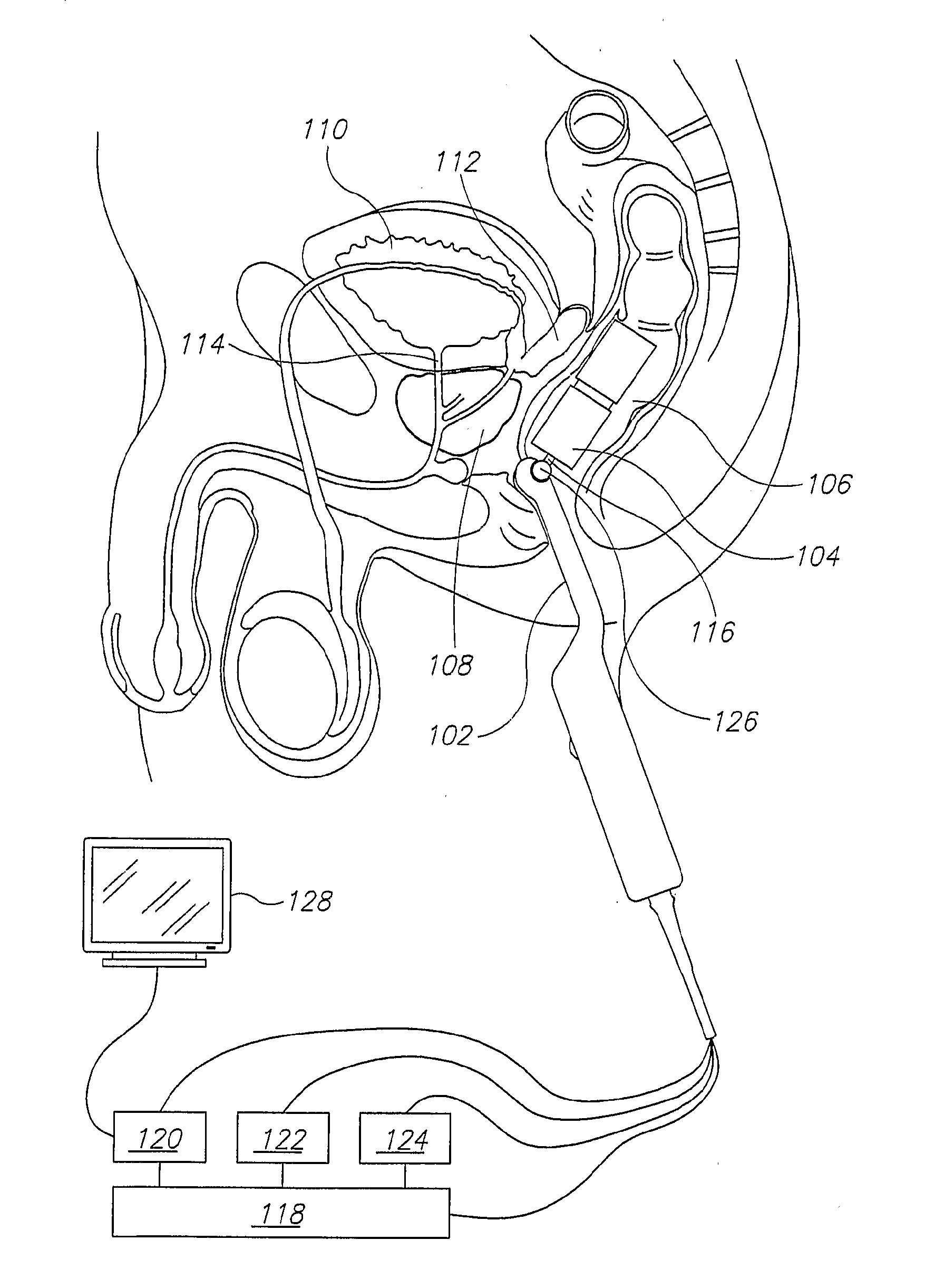
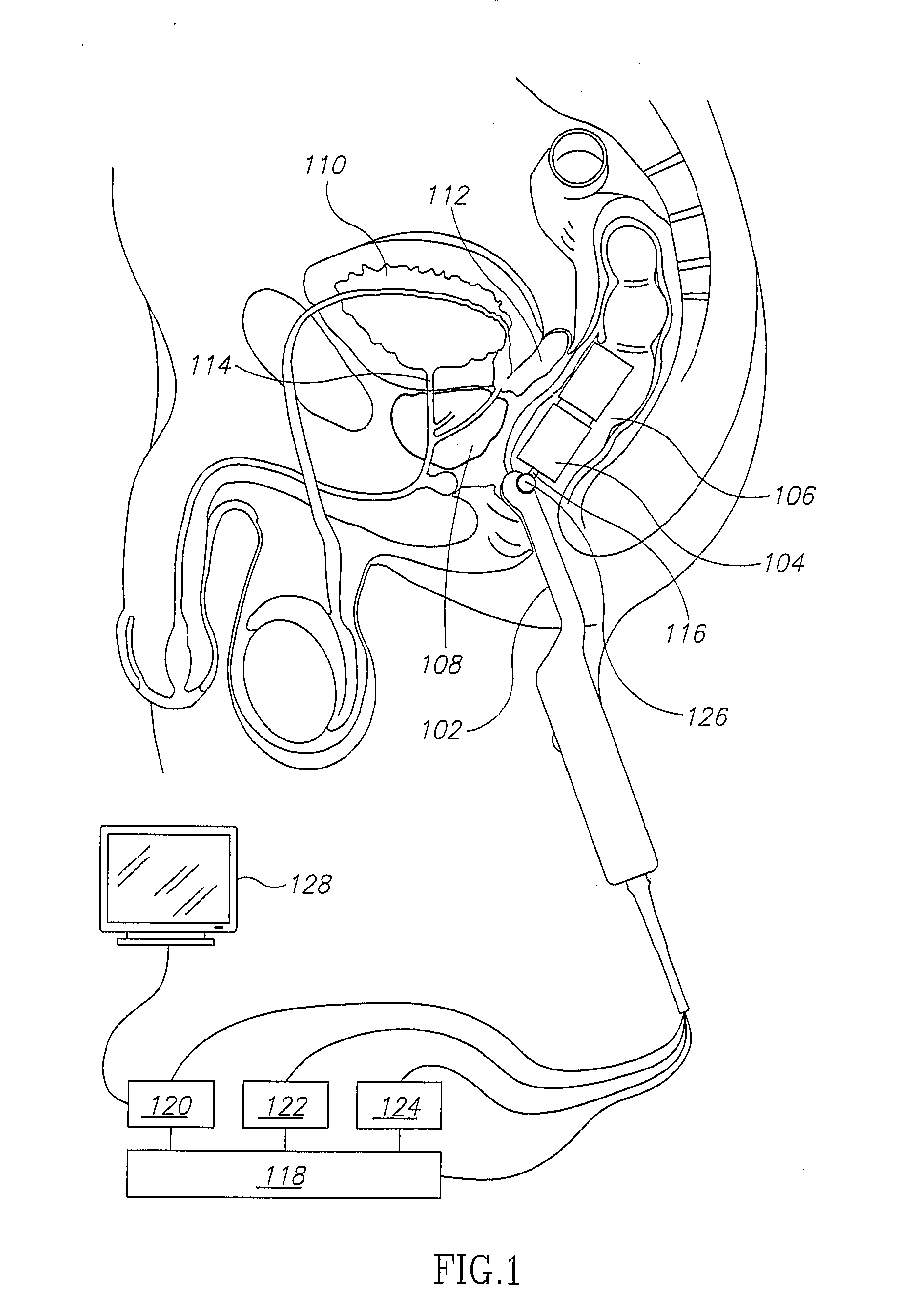
[0009]An aspect of an embodiment of the invention concerns a TMRI probe, used in the
rectum for prostate imaging. TMRI probes, because they have a highly inhomogeneous magnetic field, are more sensitive to
diffusion of protons (essentially
diffusion of water molecules) than conventional MRI, or than a Pulyer-type MRI probe which requires a relatively
homogeneous magnetic field. The increased sensitivity to diffusion optionally allows the probe to more accurately distinguish normal and malignant prostate tissue, and / or distinguish different stages of
malignancy.
[0010]In general, MRI is more sensitive to diffusion in a highly inhomogeneous magnetic field, because the excited nuclei diffuse out of
resonance more quickly. The effect of diffusion on the MRI
signal in an
inhomogeneous field is to reduce the MRI
echo signal. This is similar to the effect of the transverse
spin relaxation time, T2, which is due to diffusion in slight field inhomogeneities on a molecular scale. In conventional MRI, or even in MRI with a Pulyer-type probe, the magnetic field is so homogeneous that the reduction in
echo signal due to diffusion during a T2 time scale, is small. Hence differences in diffusion coefficient of different types of tissue within the examined sample are difficult to measure, especially when these different tissues also have different T2 values. With a highly inhomogeneous magnetic field, such as that produced by a TMRI probe, the reduction in
echo signal will, in some implementations, be dominated by diffusion effects, and not T2. This optionally enables more precise measurement of the diffusion coefficient of the various tissues, for some embodiments, at least partially independent of T2.
[0011]Potential advantages of using a TMRI probe are one or more of a) the increased ability to distinguish different types of tissue may make the probe useful in guiding biopsies or therapy; b) a TMRI probe may be used in a urologist's clinic; and c) using a TMRI probe is likely to be less expensive than using conventional MRI. Optionally, the TMRI probe is used in conjunction with a TRUS probe, which produces ultrasound images in real time, in which the boundary of the prostate is clearly visible, but in which normal and malignant tissue within the prostate are not well distinguished. By combining the MRI and ultrasound images, one can obtain an image of the prostate in real time in which the malignant regions are clearly visible. This may be useful for accurately guiding a
biopsy or a
therapeutic procedure, during which the prostate may move.
[0016]Optionally, even without an
ultrasound probe, the different MRI images are aligned, by the
software, by finding a displacement for each
MRI image which maximizes the sharpness of features in the combined image, or some other characteristic of the combined image. Optionally, the
software not only corrects for the
relative displacement of the prostate between two images acquired at different times, but also calculates the velocity of the prostate during MRI
image acquisition, and corrects for
motion artifacts in the MRI image. The velocity may be found, for example, by comparing the displacement in different images, or directly by
Doppler measurements during the
ultrasound imaging.
[0021]Optionally, the link is flexible, thereby allowing a direction of orientation of the MRI probe to vary relative to a direction of orientation of the
ultrasound probe.
