How to Maximize Graphene Oxide's Bioimaging Capabilities?
Graphene Oxide Bioimaging Background and Objectives
Graphene oxide (GO) has emerged as a promising material in the field of bioimaging due to its unique physicochemical properties. Since its discovery in 2004, researchers have been exploring its potential applications in various biomedical fields, including bioimaging. The evolution of GO-based bioimaging techniques has been driven by the need for more sensitive, specific, and non-invasive imaging modalities in medical diagnostics and research.
The primary objective of maximizing GO's bioimaging capabilities is to enhance its performance as a contrast agent and fluorescent probe. This involves improving its optical properties, biocompatibility, and targeting efficiency. Researchers aim to develop GO-based imaging agents that can provide high-resolution, real-time imaging of biological processes at the cellular and molecular levels.
One of the key trends in GO bioimaging is the development of multifunctional nanoplatforms that combine imaging capabilities with therapeutic functions. This approach, known as theranostics, allows for simultaneous diagnosis and treatment of diseases. The integration of GO with other nanomaterials, such as quantum dots or magnetic nanoparticles, is being explored to create hybrid systems with enhanced imaging properties.
Another significant trend is the modification of GO to improve its stability, reduce toxicity, and enhance its targeting abilities. Surface functionalization with biomolecules, such as antibodies or peptides, is being investigated to increase the specificity of GO-based imaging agents for particular cell types or tissues.
The advancement of GO bioimaging techniques is closely linked to progress in imaging technologies. The development of more sensitive detectors, advanced image processing algorithms, and novel imaging modalities is expected to further enhance the capabilities of GO-based imaging agents.
Researchers are also focusing on overcoming the current limitations of GO in bioimaging, such as its tendency to aggregate in biological environments and potential long-term toxicity concerns. Addressing these challenges is crucial for the clinical translation of GO-based imaging techniques.
As the field progresses, the goal is to develop GO-based imaging agents that can provide unprecedented insights into biological processes, enable early disease detection, and guide personalized treatment strategies. The ultimate aim is to create a versatile and powerful tool that can revolutionize medical imaging and contribute to improved patient outcomes.
Market Analysis for Graphene Oxide in Bioimaging
The market for graphene oxide in bioimaging is experiencing significant growth, driven by the increasing demand for advanced imaging techniques in medical diagnostics and research. Graphene oxide's unique properties, including its high surface area, excellent biocompatibility, and optical characteristics, make it an attractive material for various bioimaging applications.
The global bioimaging market is projected to reach substantial value in the coming years, with graphene oxide-based products expected to capture a growing share. This growth is fueled by the rising prevalence of chronic diseases, the need for early and accurate diagnosis, and ongoing advancements in imaging technologies. Graphene oxide's potential to enhance contrast and resolution in various imaging modalities, such as magnetic resonance imaging (MRI), computed tomography (CT), and fluorescence imaging, is a key factor driving its adoption.
In the medical field, graphene oxide-based contrast agents are gaining traction due to their ability to improve image quality and reduce the required dosage of traditional contrast agents. This not only enhances diagnostic accuracy but also minimizes potential side effects for patients. The research community is also showing increased interest in graphene oxide for cellular and molecular imaging, opening up new possibilities for studying biological processes at the nanoscale.
The pharmaceutical and biotechnology industries are major contributors to the demand for graphene oxide in bioimaging. These sectors are investing heavily in research and development to leverage graphene oxide's capabilities for drug discovery, targeted drug delivery, and theranostics. The material's potential to combine imaging and therapeutic functions in a single platform is particularly appealing for personalized medicine applications.
Geographically, North America and Europe currently lead the market for graphene oxide in bioimaging, owing to their advanced healthcare infrastructure and substantial research funding. However, the Asia-Pacific region is expected to witness the fastest growth in the coming years, driven by increasing healthcare expenditure, growing awareness of advanced imaging technologies, and rising investments in medical research.
Despite the promising outlook, challenges such as high production costs, regulatory hurdles, and the need for standardization in quality and characterization methods may impact market growth. Addressing these challenges will be crucial for realizing the full potential of graphene oxide in bioimaging applications and expanding its market penetration across various healthcare segments.
Current Challenges in Graphene Oxide Bioimaging
Despite the promising potential of graphene oxide (GO) in bioimaging applications, several significant challenges currently hinder its full utilization. One of the primary obstacles is the limited optical properties of GO, particularly its relatively low quantum yield and broad emission spectrum. These factors can result in reduced image contrast and resolution, making it difficult to achieve high-quality imaging in complex biological environments.
Another major challenge lies in the biocompatibility and toxicity of GO. While GO has shown promising results in various biomedical applications, concerns persist regarding its long-term effects on living organisms. The potential accumulation of GO in tissues and organs, as well as its interaction with cellular components, requires further investigation to ensure its safety for in vivo imaging applications.
The stability of GO in biological systems presents another significant hurdle. GO tends to aggregate in physiological conditions, which can lead to altered optical properties and reduced imaging efficiency. Additionally, the surface chemistry of GO can be affected by the complex biological milieu, potentially compromising its targeting capabilities and overall performance as a bioimaging agent.
The lack of standardization in GO synthesis and characterization methods also poses a challenge. Variations in production techniques can lead to inconsistencies in GO properties, making it difficult to compare results across different studies and hindering the development of reliable imaging protocols.
Furthermore, the challenge of targeted delivery remains a critical issue. While GO can be functionalized with various targeting moieties, achieving specific and efficient accumulation at desired sites within the body continues to be a complex task. This is particularly crucial for applications such as cancer imaging, where precise localization is essential for accurate diagnosis and treatment planning.
The development of multimodal imaging capabilities using GO is another area that requires further advancement. While GO has shown potential in various imaging modalities, integrating multiple imaging techniques (e.g., optical imaging, MRI, and PET) into a single GO-based platform remains challenging. Overcoming this hurdle could significantly enhance the diagnostic and therapeutic potential of GO in biomedical applications.
Lastly, the translation of GO-based bioimaging technologies from laboratory settings to clinical applications faces regulatory and scalability challenges. Ensuring consistent quality, developing standardized protocols, and addressing potential long-term health effects are crucial steps in bridging the gap between research and practical implementation in healthcare settings.
Existing Graphene Oxide Bioimaging Solutions
01 Graphene oxide as a bioimaging contrast agent
Graphene oxide can be used as a contrast agent for various bioimaging techniques due to its unique optical properties. It can enhance image contrast in techniques such as magnetic resonance imaging (MRI), computed tomography (CT), and fluorescence imaging. The high surface area and ability to be functionalized make graphene oxide an effective platform for developing multimodal imaging agents.- Graphene oxide as a bioimaging contrast agent: Graphene oxide exhibits excellent properties as a contrast agent for various bioimaging techniques. Its unique structure and optical properties allow for enhanced contrast in imaging modalities such as MRI, CT, and fluorescence imaging. The material can be functionalized to improve biocompatibility and targeting capabilities, making it a versatile tool for diagnostic imaging applications.
- Graphene oxide-based nanocomposites for multimodal imaging: Nanocomposites incorporating graphene oxide can be designed for multimodal imaging applications. These composites combine the advantages of graphene oxide with other imaging agents, such as magnetic nanoparticles or fluorescent dyes, to enable simultaneous imaging using multiple techniques. This approach enhances diagnostic accuracy and provides complementary information about biological systems.
- Surface functionalization of graphene oxide for targeted imaging: The surface of graphene oxide can be functionalized with various biomolecules, such as antibodies, peptides, or aptamers, to achieve targeted imaging of specific tissues or cells. This modification enhances the selectivity and specificity of graphene oxide-based imaging agents, allowing for more precise visualization of biological targets and improved diagnostic capabilities.
- Graphene oxide-based photoacoustic imaging agents: Graphene oxide demonstrates strong optical absorption properties, making it an excellent candidate for photoacoustic imaging applications. When exposed to laser light, graphene oxide generates acoustic waves that can be detected and used to create high-resolution images of biological tissues. This technique offers deep tissue penetration and high sensitivity for various biomedical imaging applications.
- Graphene oxide quantum dots for fluorescence imaging: Graphene oxide quantum dots exhibit unique fluorescence properties that can be exploited for bioimaging applications. These nanoscale particles offer advantages such as tunable emission wavelengths, high photostability, and low toxicity. Graphene oxide quantum dots can be used for long-term cell tracking, in vivo imaging, and as fluorescent probes for various biological processes.
02 Functionalization of graphene oxide for targeted imaging
Graphene oxide can be functionalized with various biomolecules, such as antibodies, peptides, or small molecules, to enable targeted imaging of specific tissues or cells. This functionalization improves the specificity and sensitivity of bioimaging applications, allowing for more accurate diagnosis and monitoring of diseases.Expand Specific Solutions03 Graphene oxide-based nanocomposites for enhanced bioimaging
Nanocomposites combining graphene oxide with other materials, such as metal nanoparticles or quantum dots, can be developed to enhance bioimaging capabilities. These hybrid materials can offer improved optical properties, increased stability, and multifunctional imaging capabilities, enabling more comprehensive and accurate diagnostic information.Expand Specific Solutions04 Photoacoustic imaging using graphene oxide
Graphene oxide exhibits strong optical absorption in the near-infrared region, making it suitable for photoacoustic imaging applications. This non-invasive imaging technique combines optical excitation with ultrasound detection, providing high-resolution images of biological tissues with enhanced contrast and depth penetration.Expand Specific Solutions05 Biocompatibility and toxicity considerations of graphene oxide in bioimaging
The biocompatibility and potential toxicity of graphene oxide are crucial factors to consider when developing bioimaging applications. Research focuses on optimizing the size, surface chemistry, and dosage of graphene oxide to minimize potential adverse effects while maintaining its imaging capabilities. Long-term studies and safety assessments are essential for clinical translation of graphene oxide-based bioimaging agents.Expand Specific Solutions
Key Players in Graphene Oxide Bioimaging
The field of maximizing graphene oxide's bioimaging capabilities is in a rapidly evolving stage, with significant market potential due to its applications in medical diagnostics and research. The global market for graphene-based products, including bioimaging applications, is projected to grow substantially. Technologically, the field is advancing quickly, with varying levels of maturity across different applications. Key players like Northwestern University, National University of Singapore, and Queen Mary University of London are at the forefront of research, developing novel techniques to enhance graphene oxide's bioimaging properties. Other institutions such as King Saud University and Indian Institutes of Technology are also contributing to the field's progression, indicating a competitive and collaborative global research landscape.
Northwestern University
National Cheng Kung University
Core Innovations in Graphene Oxide Bioimaging
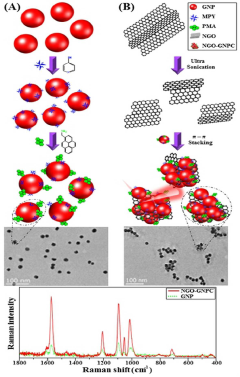
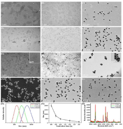
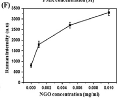
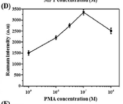
- A method for manufacturing a SERS nanoprobe with a nano-sized graphene oxide-metal nanoparticle cluster structure is developed, involving the attachment of a Raman reporter to metal nanoparticles, modification with a hydrophobic molecule, and formation of clusters through non-covalent interactions, creating highly localized plasmonic hot spots for enhanced electromagnetic fields.
- A hydro-solvo-thermal method is developed to synthesize potassium-doped graphene oxide using oak fruit as a natural source of potassium, involving solvothermal grinding, centrifugation, and nano-filtration to produce high-quality, water-soluble graphene oxide for bio-sensing and bio-imaging.
Biocompatibility and Safety Considerations
Biocompatibility and safety considerations are paramount when maximizing graphene oxide's bioimaging capabilities. The unique physicochemical properties that make graphene oxide an attractive bioimaging agent also raise concerns about its potential toxicity and long-term effects on biological systems.
One of the primary considerations is the size and shape of graphene oxide nanoparticles. Smaller particles tend to have better biocompatibility and can be more easily cleared from the body. However, they may also have reduced imaging capabilities. Striking a balance between size optimization for imaging performance and minimizing potential toxicity is crucial.
Surface functionalization plays a vital role in enhancing biocompatibility. Coating graphene oxide with biocompatible polymers or biomolecules can reduce its interactions with proteins and cells, potentially mitigating adverse effects. Additionally, surface modifications can improve stability in biological fluids and prevent aggregation, which is essential for maintaining imaging efficacy and reducing toxicity risks.
The biodistribution and clearance of graphene oxide are critical factors in assessing its safety profile. Studies have shown that graphene oxide can accumulate in various organs, particularly the liver and spleen. Understanding the kinetics of biodistribution and developing strategies to enhance clearance are essential for minimizing long-term toxicity risks.
Oxidation state and purity of graphene oxide significantly impact its biocompatibility. Highly oxidized forms tend to be more hydrophilic and less toxic, but may have reduced imaging capabilities. Conversely, less oxidized forms may offer better imaging performance but pose higher toxicity risks. Careful control of the oxidation process and thorough purification are necessary to optimize both safety and imaging efficacy.
Dose-dependent toxicity is another crucial consideration. While higher concentrations of graphene oxide may improve image quality, they also increase the risk of adverse effects. Establishing optimal dosing regimens that balance imaging performance with safety is essential for clinical translation.
Long-term effects of graphene oxide exposure remain a concern. Chronic toxicity studies are needed to assess potential carcinogenicity, immunogenicity, and reproductive toxicity. Additionally, the potential for graphene oxide to cross biological barriers, such as the blood-brain barrier, requires careful evaluation to ensure safety in various applications.
In conclusion, maximizing graphene oxide's bioimaging capabilities while ensuring biocompatibility and safety requires a multifaceted approach. Ongoing research and rigorous safety assessments are essential to unlock the full potential of graphene oxide in bioimaging applications while minimizing risks to human health and the environment.
Regulatory Landscape for Nanomaterials in Bioimaging
The regulatory landscape for nanomaterials in bioimaging is complex and evolving, reflecting the rapid advancements in nanotechnology and its applications in medical imaging. Regulatory bodies worldwide are grappling with the unique challenges posed by nanomaterials, particularly in the context of their use in biological systems.
In the United States, the Food and Drug Administration (FDA) plays a central role in regulating nanomaterials for bioimaging applications. The FDA has established specific guidelines for the evaluation of nanomaterials in medical products, including those used in imaging. These guidelines address issues such as characterization, toxicity assessment, and quality control of nanomaterials.
The European Union has implemented the REACH (Registration, Evaluation, Authorization, and Restriction of Chemicals) regulation, which applies to nanomaterials used in various applications, including bioimaging. Additionally, the European Medicines Agency (EMA) has developed specific guidance for nanomedicines, which encompasses imaging agents based on nanomaterials.
In Asia, countries like Japan and South Korea have also established regulatory frameworks for nanomaterials. The Japanese government has implemented the "Guidelines for Ensuring the Safety of Nanomaterials" which cover various aspects of nanomaterial use, including in medical applications.
A key challenge in the regulatory landscape is the lack of standardized testing protocols for nanomaterials. Efforts are underway to develop internationally recognized standards through organizations like the International Organization for Standardization (ISO) and the Organisation for Economic Co-operation and Development (OECD).
Regulatory bodies are particularly concerned with the potential long-term effects of nanomaterials on human health and the environment. As a result, there is an increasing emphasis on lifecycle assessments of nanomaterials used in bioimaging, from production to disposal.
The regulatory landscape also reflects a growing focus on the ethical implications of nanomaterial use in medical imaging. This includes considerations of patient privacy, informed consent, and the potential for unintended consequences of long-term exposure to nanomaterials.
As research into graphene oxide's bioimaging capabilities continues to advance, regulatory frameworks are likely to evolve. This may include the development of specific guidelines for graphene-based materials in medical imaging, addressing their unique properties and potential risks.